
A quick guide for morphometric fingerprint analyses
How to compare the shape of an object with that of another one? Purely "qualitative" morphological descriptions might be an easy option. Statements like "clasts from sample A are more rounded than those of sample B" or "sample C ash comprises mossy shaped grains, while ash from sample D is characterized by elongated tubular surface features" are found in basically any article reporting fieldwork results.
This kind of description is somewhat subjective, which is the reason why more quantitative approaches have been developed [1]. The aim of such systems was to use sets of quantitative shape descriptors, which unambiguously describes the outlines of an object. Describing the shape of an object is called "morphometry", which is why we also talk about "morphometric analyses" and "morphometric systems".
In general, we distinguish at least three morphometric approaches used in volcanology [2]:
- 2D shape analysis using projected silhouettes: the grains are imaged e.g., by an optical or electron microscope mounted on carbon tape; then the areas of these objects are projected on a plane. This method has a couple of advantages and basically any automatic shape analyzer on the market uses it. (These are machines, where you fill in your sample, and they spit out the results). But even if you do not have such a fancy machine, doing 2D shape analysis is quite an easy task and requires only little training. The equipment you need (binocular microscope, or scanning electron microscope) should be available at most research facilities. The disadvantage of this method is that it does not reveal anything about the particle's interior. This might be a problem, if you try to distinguish e.g. juvenile from lithic clasts.
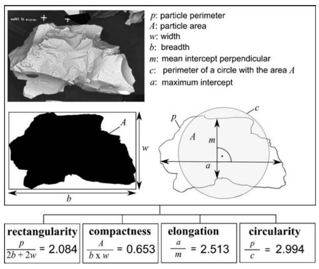
2D shape analysis using silhouette. In this example, the shape of the depicted ash grain is quantified by four parameters, based on some basic geometric measures: rectangularity, compactness, elongation and circularity. This system is also known as "image particle analysis" or IPA [3] (Figure adapted from Dürig et al. (2012) [4])
- 2D shape analysis, using cross sections: for this method you have to prepare either polished epoxy grains or make thin sections. The advantage is that you can also see the grain interior and use the samples to simultaneously analyze (2D) vesicularity and crystallinity. The disadvantages are, however, a more complicated preparation process and therefore a larger temporal effort. Furthermore the polishing step adds a degree of uncertainty, due to the fact that there are (quasi) infinite possibilities for how the cross-section is located within the grain. Although there are often filter processes applied to limit it, the number of degrees of freedom is larger than those for the 2D silhouette method. Statistically, this means that we expect that larger sample sizes (higher number of particles) are required to get representative results, compared to the silhouette method. This further increases the temporal effort of the cross-section method.
Importantly (and unfortunately), results obtained by 2D cross-section method cannot be compared to those obtained based on silhouettes [1, 2].
- - 3D analysis by X-ray micro-tomography: The particles are scanned in 3 dimensions, which means that it provides the most detailed description of grains. The advantages towards the 2D methods are obvious: both, surface morphology and internal structures can be quantified (including the "actual" (3D) vesicularity and crystallinity!). And the resulting 3D images are just super-cool!
The reason, why 3D is still barely applied is simply that these machines are very expensive (and often involve high running costs), scanning at high resolution requires training and/or experienced staff, and takes quite some time. Considerable temporal effort is also required for particle mounting, image correction and reconstruction, particle image segmentation in 3D and conducting the shape analysis itself. Availability, time and potentially high costs are therefore the three factors which limit the use of this method.
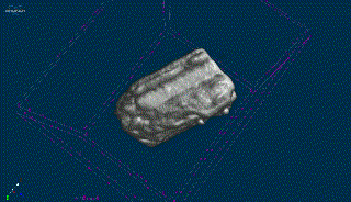
3D scan of ash grain retrieved at Havre volcano by applying X-ray microtomography. Note the clearly visible tubular structure.This scan was conducted at University of Bari, Italy.
In the past, basically every research group used their own morphometric system. This resulted in a somewhat "Babylonian confusion" with published results that are only of limited use: to reproduce them you have to use the same system.
In an effort to put this unpleasant state of anarchy to an end, recently, an inter-lab effort was made to introduce a standardized protocol for particle shape analysis [2, 5].
As a first step towards unification, a freeware named PARTISAN (particle shape analyzer) was developed for Matlab, which computes 24 shape parameters from 5 different morphometric systems [1], thus serving as "Rosetta stone" in morphometry.
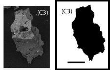
Ash particle and its silhouette. (Adapted from Dürig et al. (2018) [1])
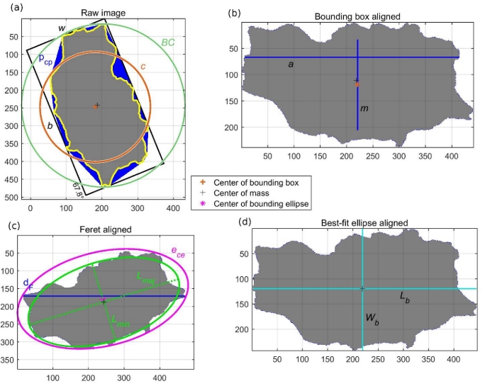
Figure produced by PARTISAN. The indicated measures are used to produce 24 shape factors, used in five different morphometric systems. (Figure adapted from Dürig et al. (2019) [1])
Next to presenting a graphical display, PARTISAN stores the resulting parameters in a .csv table for further statistical evaluation. To help volcanologists with that, a software has been specifically designed to conduct all kind of statistical tests using the PARTISAN output. This programm is called DendroScan [6], and it can be downloaded here.
Besides that, researchers can fall back on a huge variety of additional statistical test methods, which have all their pros and cons, and are described in detail in a recently published review article [7].
When comparing different samples, a typical goal is to find out if the compared samples are morphometrically distinguishable.
Another frequent quest is to find out which of a set of samples is most similar to a certain reference sample [2, 4]. Typically, the latter strategy is the one used by us "experimental volcanologists": in order to find out how volcanic ash samples retrieved at the volcano were produced, we conduct a variety of different fragmentation experiments, collect the resulting fragments and finally compare their shapes with the "natural" sample.
This "reconstruction" approach is therefore very much like you might know it from the CSI crime show. It is this strategy we also followed to solve the Havre ash mystery.
-> Lab-scaled melt fragmentation experiments
-> Fingerprints of the Havre eruption
<- Why fine volcanic ash particles are good eye-witnesses
Annotations:
The Havre ash project was funded by a Marsden grant, New Zealand.
The lines of thought above are based on considerations detailed in Dürig et al. (2019) [1] and Dürig et al. (2012) [2], if not cited otherwise. Please let me know if you have any comment or suggestions for changes. My email address is: tobi[_at]hi.is
References and Annotations
[1]: Dürig, T., Bowman, M.H., White, J.D.L., Murch, A., Mele, D., Verolino, A., Dellino P. (2019): PARTIcle Shape ANalyzer PARTISAN – an open source tool for multi-standard two-dimensional particle morphometry analysis. Annals of Geophysics 61 (6), 671.
DOI: 10.4401/ag-7865
[2]: Ross P.-S., et al. (2022): Standardized analysis of juvenile pyroclasts in comparative studies of primary magma fragmentation; 1. Overview and workflow. Bulletin of Volcanology 84(1): 13. doi: 10.1007/s00445-021-01516-6
[3]: P. Dellino and L. La Volpe (1996): Image processing analysis in reconstructing fragmentation and transportation mechanisms of pyroclastic deposits. The case of Monte Pilato-Rocche Rosse eruptions, Lipari (Aeolian islands, Italy). Journal of Volcanology and Geothermal Research 71 (1), 13-29.
[4]: Dürig, T., Mele, D., Dellino, P., Zimanowski, B. (2012): Comparative analyses of glass fragments from brittle fracture experiments and volcanic ash particles. Bulletin of Volcanology 74 (3), 691-704.
[5]: Comida P.P., et al. (2022): Standardized analysis of juvenile pyroclasts in comparative studies of primary magma fragmentation; 2. Choice of size fraction
and method optimization for particle cross‑sections. Bulletin of Volcanology 84(1): 14. doi: 10.1007/s00445-021-01517-5
[6]: Dürig T., Schmidt, L.S., White J.D.L., Bowman, H.M. (2020): DendroScan – an open source tool to conduct comparative statistical tests and dendrogrammatic analyses on particle morphometry. Science Reports 10:21682.
DOI: 10.1038/s41598-020-78698-0
[7]: Dürig T., Ross P.-S., Dellino P., White J.D.L., Mele D., Comida P.P. (2021): A review of statistical tools for morphometric analysis of juvenile pyroclasts. Bulletin of Volcanology 83(1): 79. doi: 10.1007/s00445-021-01500-0